 BIOGEOSPHERE
Bilingual Biology and Geology
I.E.S. "J.S.Elcano (Sanlúcar Bda.)
BIOGEOSPHERE
Bilingual Biology and Geology
I.E.S. "J.S.Elcano (Sanlúcar Bda.)
2.2. The heart
The heart is a muscular organ about the size of a fist. It is located in the middle of thoracic cavity, slightly left side and between the lungs. Its function is to pump the blood to the whole body.
The heart wall is formed by three layers:
- Pericardium: It is the most external layer. It is a connective tissue layer
that joins to the heart:
- The nerves that innerve the heart (responsible for the impulse
that makes the heart beats)
- The blood vessels that irrigate the heart (coronary artery and vein)
- Myocardium: It is in the middle. It is a thick layer of cardiac muscle
tissue.
- Endocardium: It is the most internal layer. It is a connective tissue layer
that lines the inner cavities of the heart and made up the valves.
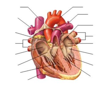
The heart is divided into two parts, left and right, by the interventricular septum. These two parts are not communicated.
Each part of the heart is divided into two chambers:
- Atrium: It is the upper cavity. It is smaller and its walls are thin and elastic.
- Ventricle: It is the lower cavity. It is bigger and its walls are thick and strong.
The two chambers of the same side are communicated by a valve, which allows the pass of blood from atrium to ventricle but not in inverse way.
- Bicuspid (mitral) Valve: It is in the left side. It is formed by two languets.
- Tricuspid Valve: It is in the right side. It is formed by three languets.
The veins arrive to atria and bring blood back to the heart:
- Pulmonary Veins arrive to the left atrium (2 come from the left lung
and 2 from the right lung)
- Inferior Vena Cava (brings blood from the inferior part of body)
and Superior Vena Cava (brings blood from the superior part of body)
arrive to the right atrium.
Arteries exit from ventricles and carry blood to the lungs and the rest of body:
- Pulmonary Artery exits from right ventricle and later divides into two branches,
one for each lung.
- Aorta Artery exits from left ventricle and carries blood to all the body.
The blood exiting from ventricles is controlled by Sigmoid Valves located
at the beginning of arteries. These valves prevent that blood goes back to ventricles.
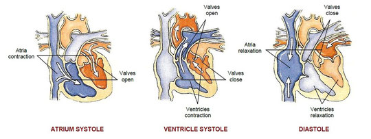
a) Cardiac cycle (heartbeat)
The cardiac cycle is the join of phases which heart pass through in each beat.
1st) Atrium systole (atrium contraction)
- Atria, full of blood, contract.
- Mitral and tricuspid valves open (because atrium pressure is bigger than ventricle pressure)
- Blood passes to ventricle
- Mitral and tricuspid valves close (because atrium pressure is smaller than ventricle pressure)
2nd) Ventricle systole (ventricle contraction)
- Ventricles, full of blood, contract.
- Sigmoid valves open (because ventricle pressure is bigger than artery pressure)
- Blood passes to arteries
- Sigmoid valves close (because ventricle pressure is smaller than artery pressure)
3rd) General diastole (heart relaxation)
- Atria and ventricles, empty of blood, relax.
- Blood pass to atria from the veins by suction.
b) Cardiac parameters
- Heart rate
It is the number of times heart beats by unit of time. Usually it is about 70 beats/min, but it depends on age, sex and size. It also changes to adapt to physiologic demands. It rises during exercise or exciting situations and falls during rest or sleep.
- Cardiac output
It is the volume of blood which heart can pump in a minute.
Usually it is about 5.5 litres/min, but is larger in sport people because their heart is bigger.
- Stroke volume
It is the volume of blood which heart can pump in each beat.

ACTIVITIES
After reading the text, copy and answer the following questions into your notebook:
Remember: you must make complete sentences.
2.6. Could the heart be a non-muscular organ? And a non-hollow organ? Why?
2.7. Name the labelled parts of the picture with the terms used in the text.
(Remember, in anatomic sketches left and right sides are inversed)
2.8. Listen and indicate what part of the heart is described:
a. Ventricles
b. Atria
c. Tricuspid valve
d. Bicuspid (Mitral) valve
e. Sigmoid valves
2.9. How is it assured that the flow of blood within the heart is always
unidirectional?
Vein -> Atrium -> Ventricle -> Artery
2.10. Calculate the beat volume of a sport-man which cardiac frequency
is 40 beats/min and which cardiac waste is 6.5 litres/min.
After an intensive exercise his cardiac frequency is 120 beats/min,
how have the other parameters changed?


(Diccionario Ing-Esp)
(Juegos de vocabulario)
(Visual dictionary)
(Visual dictionary)
(Glosario de C. Naturales)
(Science audio-glossary)
(Science audio-glossary)
(Pronunciación)
(Enciclopedia de C. Naturales)
(Tabla periódica)

Eva Mª
López Rodríguez
Departamento
Biología y Geología
IES " J. S. Elcano"
Sanlúcar de Barrameda